柔道整復師のための解剖学シリーズ 顎関節1
いよいよ顎関節脱臼のことについて触れてゆくことにしましょう。関節包が一部明確でない1)、外側靭帯など靭帯構造が明確でない…などの解剖学的特徴をもつヒトの顎関節が脱臼するとなると、いわゆる関節頭が生理的範囲を越えて、関節窩を逸脱して、靭帯が断裂して…などの脱臼の定義をどこに当てはめられるでしょうか。
Fig. 1 のような関節包 2) は、ヒト顎関節には存在しないと考えるべきです。 では顎関節全体を包む膜様の組織はないのでしょうか?、Masticator space fasciaと呼ばれる臨床上重要な役割をもつ膜が知られています。この膜は放射線診断学において腫瘍の転移を知る上で、とても重要な組織として捕えられています 3)。顎関節はこのスペースの中にあるといってよいのです。
しかし、顎関節内側後方には円板後方静脈叢があることはすでに述べたことです。Bilaminar zone との呼称されるここでは、この膜の連続性はありません。また、円板の前方は外側翼突筋が停止しています。
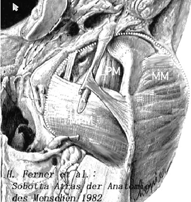
円板の外側に対する筋の停止については議論がなされているようですが、筆者が検体で観察した限りでは、どの関節でも円板の外側では側頭筋や咬筋の線維が停止していました。おそらくこれらの筋が、右顎関節を下方から見ている。点線のラインにMasticator Space Fascia と呼ばれる膜が停止します。顎関節はこの膜の中にあるといってよいでしょう。咀嚼筋の活動による筋血流の影響を受けることが理解できます。微妙な顎関節の動きに追従する円板の生理的な動きを調節していると考えられます。つまりヒト顎関節後外側には Gray’s Anatomy 37 th 4) に示されるような関節包は存在しないと考えられます。・・・・・以後割愛


参考文献: 1,Yosuke Shiraishi et al.: A new retinacular ligament and vein in the posterolateral articular disk in human temporomandibular joint. Clin Anat, 1995; 8: 3, 208-213. 2. Wilkes C.H.: Arthrography of the temporomandibular joint. Minnesota Med. 1978, 61; 645. 3, Curtin H.D.: Separation of the masticator space from the perapharyngeal space. Radiology, 1987, 163; 1, 195-204. 4, Williams et al. (eds): Arthrology, Gray’s Anatomy 37th ed. New York, Churchill Livingstone, p 485-489, 1989.5, Miyake M. et al.: morphological study of the human maxillofacial venous vasculature; examination of venous valves using the corrosion resin cast technique. Anat Record 1996; 244, 126-132. 6, Kai S. et al.: Clinical symptoms of open lock position of the condyle. Relation to anterior dislocation of the temporomandibular joint. Oral Surg Oral Med Oral Pathol. 1991, 74; 2 143-148. 7, Nitzan D.W.: Temporomandibular joint “open lock” versus condylar displacement: signs and symptoms, imaging, treatment, and pathogenesis. J Oral Maxillofac Surg. 2002, 60; 5 56-511.
